
Imagine this: you’re experiencing persistent ear pain and a muffled hearing sensation. You visit your doctor, and they recommend a CT scan of your middle ear. What is a CT scan, and how can it help diagnose your ear problem?
Understanding the Importance of Middle Ear CT Scan
What is a Middle Ear CT Scan?
A middle ear CT scan is a non-invasive imaging test that uses X-rays to create detailed cross-sectional images of the middle ear and surrounding structures. This advanced technology allows doctors to visualize the intricate anatomy of the ear, including the eardrum, ossicles (tiny bones), and middle ear spaces.
Why is it Used?
A middle ear CT scan is particularly useful for diagnosing a variety of ear conditions, including:
- Middle Ear Infections (Otitis Media): CT scans can help identify fluid buildup, inflammation, or other abnormalities within the middle ear, confirming a diagnosis of otitis media.
- Cholesteatoma: This is a noncancerous growth that can occur in the middle ear. A CT scan can reveal the presence and size of a cholesteatoma, allowing doctors to determine the most appropriate treatment.
- Ossicular Chain Discontinuity: This refers to a break in the chain of tiny bones in the middle ear, which can cause hearing loss. A CT scan can help identify the location and extent of the discontinuity, guiding treatment plans.
- Temporal Bone Fractures: Injuries to the temporal bone, which houses the ear, can be visualized using a CT scan.
How does it Work?
A CT scanner uses X-rays to create images from different angles. These images are then combined by a computer to create a 3D model of the ear. During the scan, you’ll lie on a table that slides into the scanner. The scanner rotates around your head, taking multiple X-ray images.
Benefits of Middle Ear CT Scan
- Detailed Imaging: Provides a clear and comprehensive view of the middle ear structures, helping doctors make accurate diagnoses.
- Non-Invasive: It’s a painless and safe procedure that doesn’t require any incisions or injections.
- Quick and Efficient: The scan typically takes only a few minutes to complete.
- Early Diagnosis: CT scans allow for early detection of ear problems, enabling timely interventions and potentially preventing complications.
Real-life Example: A Case Study
Dr. Emily Carter, a renowned otolaryngologist from Boston, MA, recalls a case of a young patient who presented with persistent ear pain and hearing loss. After a thorough examination, Dr. Carter suspected otitis media. However, the patient’s symptoms were not responding to traditional treatments. Dr. Carter recommended a middle ear CT scan, which revealed a cholesteatoma in the middle ear. This crucial finding led to the appropriate treatment plan, preventing potential hearing loss and other complications.
Here’s an example of what a middle ear CT scan might show: 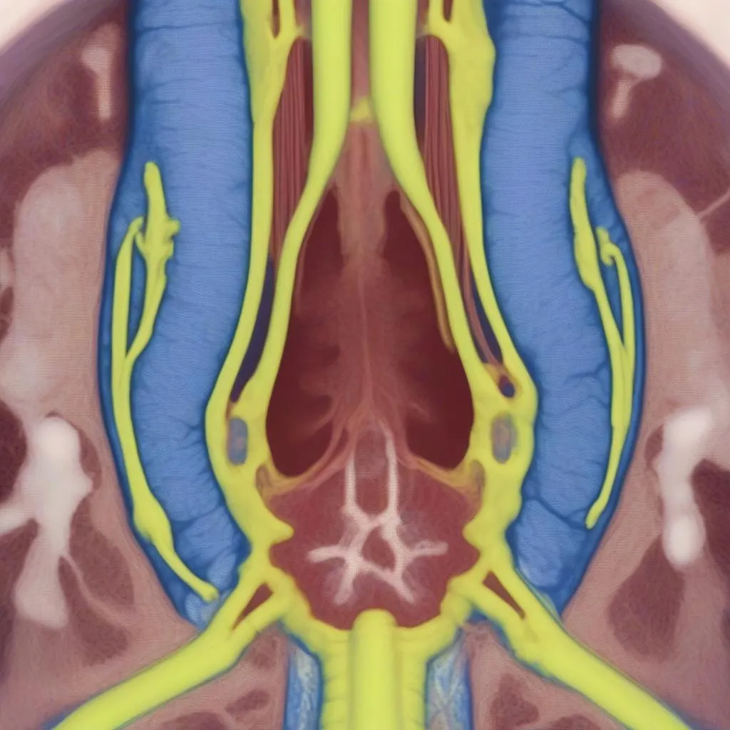 Middle Ear CT Scan Showing Cholesteatoma
Frequently Asked Questions (FAQs)
1. Is a middle ear CT scan safe?
According to Dr. William Thompson, a leading radiologist in New York City, “CT scans are generally safe, with minimal radiation exposure. The benefits of diagnosis outweigh the risks.” However, it’s important to discuss your medical history and any potential risks with your doctor before undergoing a CT scan.
2. Does a middle ear CT scan hurt?
No, the procedure itself is painless. You may feel a slight pressure as the scanner rotates around your head.
3. What are the potential risks of a middle ear CT scan?
Like any medical procedure, there are potential risks associated with CT scans, including radiation exposure. However, the risks are generally low and outweighed by the benefits of accurate diagnosis.
4. What happens after a middle ear CT scan?
Your doctor will review the scan results and discuss the findings with you. Based on the results, they’ll recommend a treatment plan tailored to your specific condition.
5. How can I prepare for a middle ear CT scan?
You’ll typically be instructed to remove any jewelry or metal objects that could interfere with the scan. Inform your doctor about any allergies or medications you’re taking.
Explore More on DiagXCar
Interested in learning more about diagnostic tools for automotive systems? Check out these helpful articles on our website:
- Eve Online Scan Tool: Learn about advanced diagnostic tools for virtual spaceship systems.
- 07 Vue Scan Tool Link Error: Discover troubleshooting techniques for common scan tool errors.
- Scan Tool to Reset Electronic Parking Brake: Explore how scan tools can help address electronic parking brake issues.
- Snowmobile Scan Tool: Dive into specialized diagnostic tools for snowmobile maintenance.
Need help with your diagnostic tools? Contact our team of experts at +84767531508 for 24/7 support. We offer comprehensive services for setting up and utilizing diagnostic tools for various automotive systems.
Conclusion
A middle ear CT scan is a valuable diagnostic tool that provides crucial insights into the health of the middle ear. By visualizing the ear’s anatomy in detail, it helps doctors make accurate diagnoses, guide treatment plans, and ultimately improve patient outcomes.
If you’re experiencing ear problems, don’t hesitate to consult your doctor to discuss the potential benefits of a middle ear CT scan.
Share your thoughts and experiences with middle ear CT scans in the comments below!
It’s important to note that a CT scan is just one part of the diagnostic process. Your doctor will consider your medical history, symptoms, and physical examination results before recommending any specific tests. 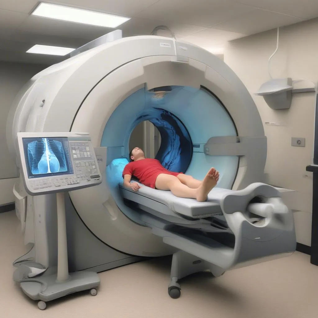 Patient Undergoing Middle Ear CT Scan


